Lab-grown, “living” heart tissues: a major breakthrough

By UdeMnouvelles
In 5 seconds Described as “hearts on a chip,” the engineered tissues are produced through 3D bioprinting with a bio-ink developed in a laboratory at the UdeM-affilated CHU Sainte-Justine.
The researchers' approach more accurately reflects the complexity of the human myocardium.

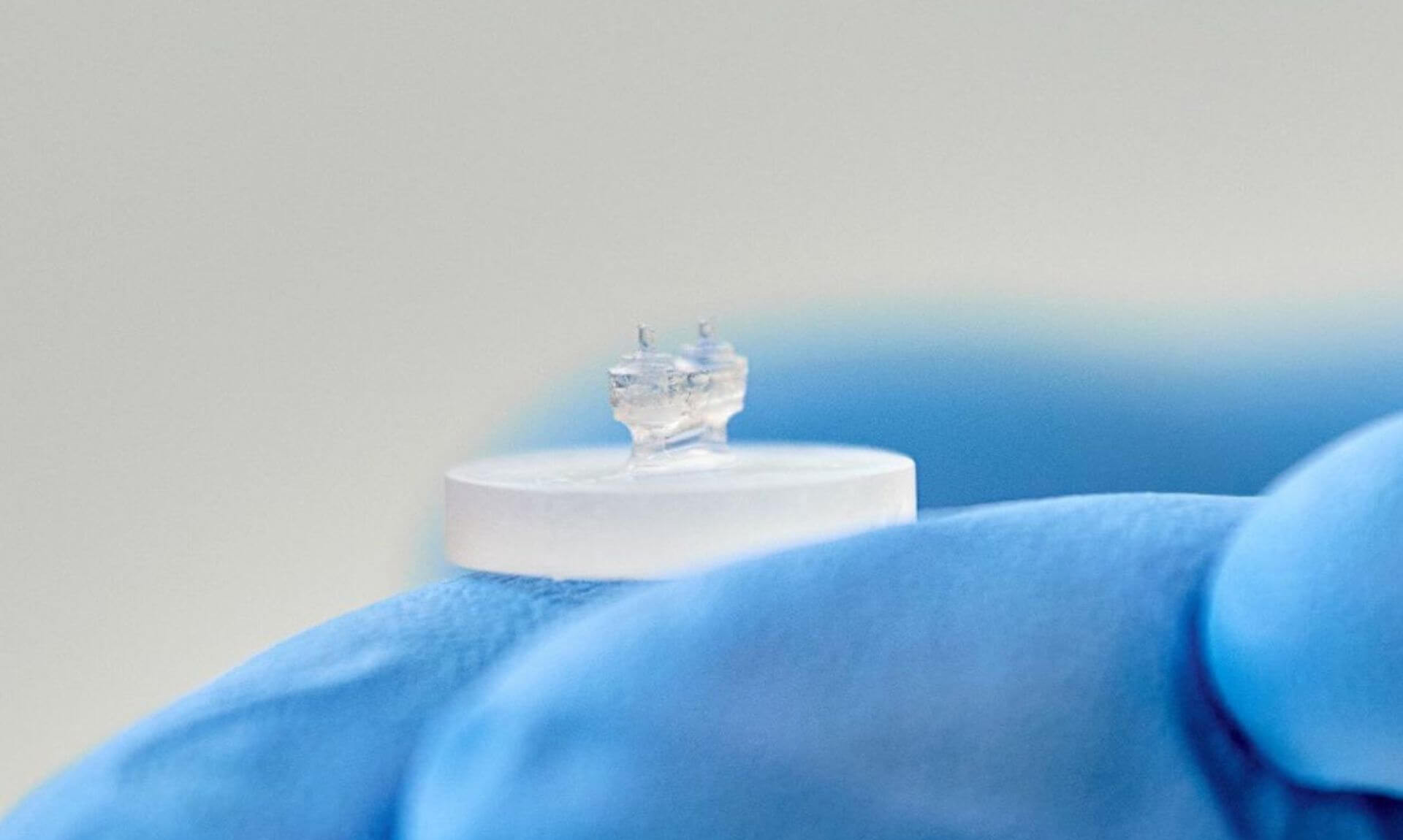
Scientists at Université de Montréal and its affiliated Centre de recherche Azrieli du CHU Sainte-Justine have made a major advance in their research into cardiovascular disease: they've created a functional, three-dimensional heart tissue that can beat autonomously in vitro.

The tissue incorporates micro-sensors that make it possible to do finely tuned, real-time analysis of its contractile properties. This advance marks an important step forward for modeling human cardiac diseases and conducting preclinical drug testing.

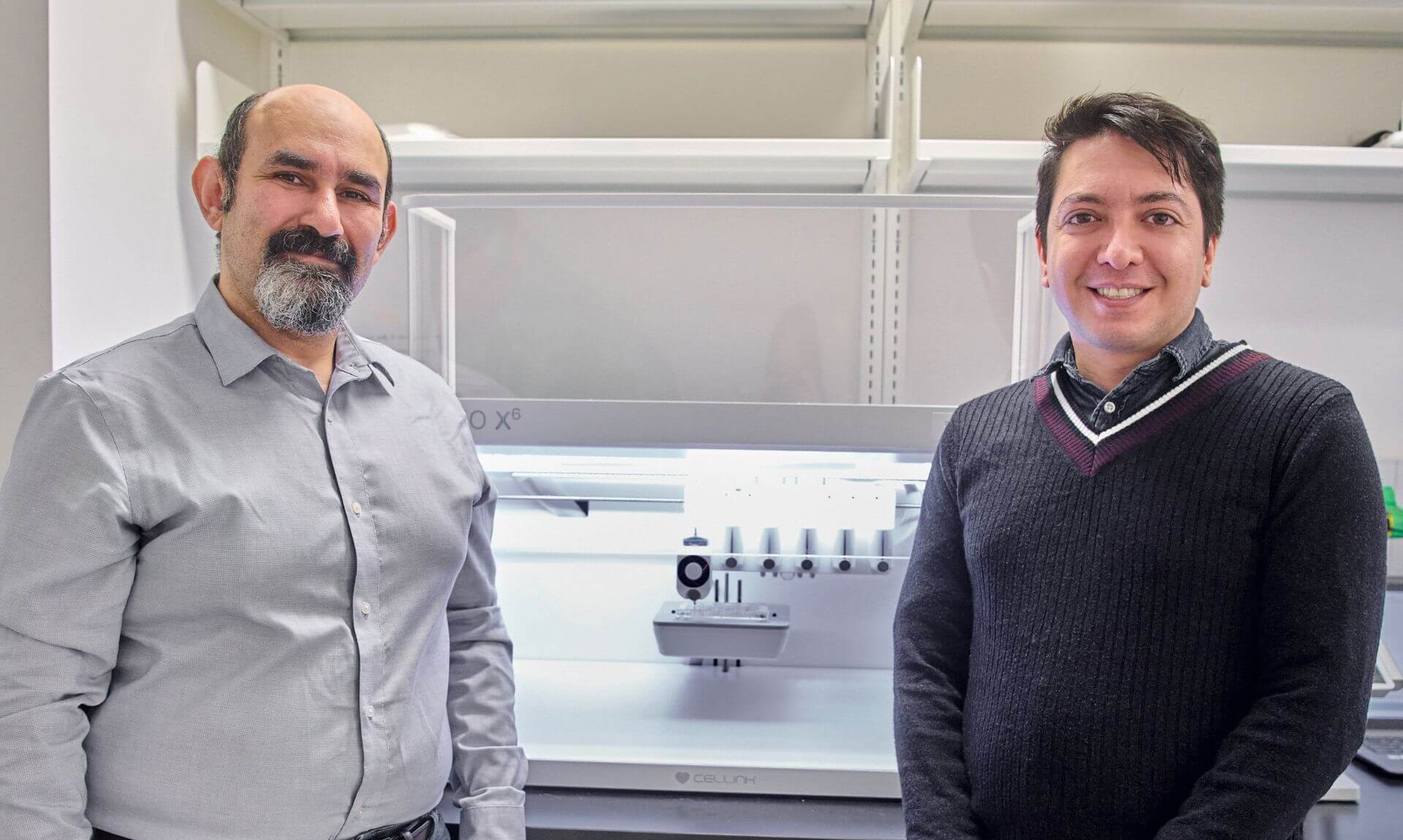
Led by UdeM pharmacology and physiology professor Houman Savoji at his laboratory at CHU Sainte-Justine, along with UdeM PhD student Ali Mousavi, the research is detailed in a study published in the scientific journal Nano Micro Small.

'Hearts on a chip'

Described as “hearts on a chip,” the engineered heart tissues are produced through 3D bioprinting with a bio-ink developed in Savoji’s laboratory using patient-harvested stem cells, enabling personalized human heart models to be created.

An initial version of the technology was publicized two years ago, in a study in Applied Materials Today.

The new study marks a major step forward: the direct integration of ultra-soft, biocompatible and fluorescent mechanical sensors within the heart tissue itself. These sensors allow for unprecedented precision in measuring the contractile forces generated at both the cellular level and across the entire tissue, using non-destructive optical methods.

Unlike existing “heart-on-a-chip” platforms—often limited in their ability to capture localized forces within dynamic 3D tissues—this approach delivers high-resolution, real-time, mechanical data. It therefore more accurately reflects the complexity of the human myocardium, the muscle responsible for cardiac contraction.

The researchers also measured calcium activity within the tissues, visualizing in real time the calcium waves that trigger each heartbeat. They further demonstrated that their “hearts on a chip” respond to drugs just like real cardiac tissues, confirming the model’s sensitivity for pharmacological screening.

Next up: models for more diseases

The team is now working to develop models of cardiovascular diseases such as dilated cardiomyopathy and some arrhythmias, by comparing tissues derived from the cells of patients living with these conditions to tissues generated from those of healthy individuals.

Ultimately, this technology could enable the modeling of a wide range of cardiac disorders and the precise assessment of potential therapies, the researchers say.

“The ability to observe the tissue’s response to different compounds in real time represents a major advantage for preclinical development and translational research,” said Mousavi, the study's first author.

“This allows us to test directly on a patient’s own cells, without any invasive procedures.”

Added Savoji, the study's principal investigator: “This breakthrough brings us even closer to true precision health, by giving us the ability to identify the most effective medication for each person before treatment is even administered."

Share

Media requests

CHU Sainte-Justine
Université de Montréal
Phone: 514-343-6111, ext. 67960